STM Observation of Self-Assembled Layers of 10,12-Pentacosadiyn-1-ol
Physisorbed on Graphite

Self-assembled layers of 10,12-pentacosadiyn-1-ol (PCDYol) physisorbed on graphite have been investigated by Scanning Tunneling Microscopy (STM). In STM images, a herringbone molecular arrangement was observed as in the case of other alkanols. Parallel bright lines, which are assigned to the columns of the diacetylene parts were observed. Among the lines, two different levels of brightness appearing in an alternating manner were observed. This is due to the difference in orientation of the alkyl chains with respect to the graphite surface lattice.

(by K. Noda & D. Takajo)

Fig. 1

Fig. 1. Molecular structure of PCDYol.

Fig. 2

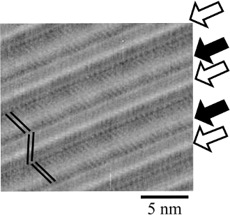
Fig. 2. A typical high resolution image of adlayer. Arrows on the right side indicate two levels of brightness of the lines of diacetylene moieties. White ones mean bright lines and black ones. The bars on the left side indicate the alkyl chain directions.

Fig. 3

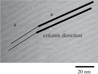
Fig. 3. An STM image at a domain boundary. Difference in line thickness means that of the column directions.

Fig. 4

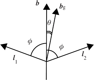
Fig. 4. Sketch of the vectors discussed. For clarity, θ is emphasised and length of vectors is not precise.

Copyright © Research Center for Structural Thermodynamics, Graduate School of Science, Osaka University. All rights reserved.